Exploring the Frontiers of Optical Medical Imaging
Exploring the Frontiers of Optical Medical Imaging
Optical imaging technologies are redefining the way healthcare professionals visualize and understand the human body. From early-stage diagnostics to real-time surgical guidance and advanced biomedical research, optical techniques are enabling remarkable insights into physiological and molecular processes.
In this overview, we’ll introduce two foundational approaches in optical imaging—diffusive and ballistic optical imaging—and also touch on hybrid and nuclear imaging methods like photoacoustic imaging and positron emission tomography (PET), all of which are transforming medical diagnostics and research.
Diffusive Optical Imaging (DOI)
Diffusive optical imaging uses near-infrared (NIR) light, often through spectroscopy or fluorescence, to probe the optical characteristics of soft biological tissues. Because soft tissue diffuses light heavily, DOI is particularly effective for monitoring metabolic activity and changes in chromophores like hemoglobin or cytochromes.
Time-resolved systems, which deliver picosecond laser pulses and capture the exiting light using time-correlated single photon counting (TCSPC), provide data that can be computationally reconstructed into optical images. These techniques are invaluable for applications such as breast cancer screening, cerebral hemodynamics, muscle metabolism, and more.
When DOI is extended to 3D imaging, it becomes diffusive optical tomography (DOT), which continues to gain ground in both research and clinical environments.
Ballistic Optical Imaging
In contrast to diffusive techniques, ballistic optical imaging relies on detecting photons that pass through tissue with minimal scattering—so-called “ballistic photons.” These unscattered photons retain more spatial resolution and are critical for producing crisp, high-resolution images.
Techniques like optical coherence tomography (OCT) and ultrafast gated ballistic scanning systems fall under this category. While limited in penetration depth, ballistic imaging excels in clarity and is often favored in applications requiring fine detail, such as ophthalmology or dermatology. Advanced image reconstruction algorithms can extend its utility deeper into tissue.
Photoacoustic Imaging
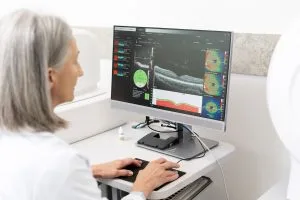
Photoacoustic imaging (or optoacoustic imaging) merges the strengths of optical imaging and ultrasound. A pulsed laser induces thermoelastic expansion in tissues, which generates ultrasound waves detected by transducers. This hybrid technique captures both structural and functional information with impressive resolution and depth.
Non-ionizing and suitable for both endogenous and exogenous contrast agents, photoacoustic imaging is advancing rapidly with techniques such as photoacoustic tomography (PAT) and photoacoustic microscopy (PAM) finding applications in oncology, neurology, and cardiovascular imaging.
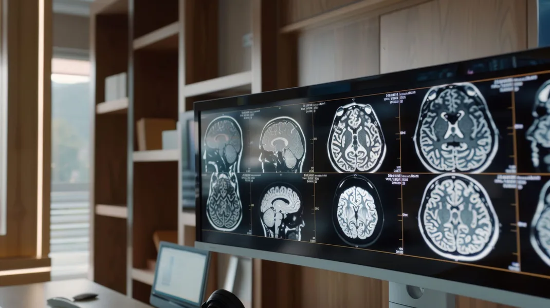
Positron Emission Tomography (PET)
While not an optical method, PET complements optical imaging by providing metabolic and molecular data that other imaging techniques might miss. It involves the injection of a radioactive tracer, which highlights areas of high metabolic activity—often associated with tumors or diseased tissues.
PET scans are frequently paired with CT or MRI to deliver high-resolution, multimodal images. Despite requiring specialized facilities and safety precautions, PET remains a cornerstone of advanced diagnostic imaging, especially in oncology and neurology.
Custom Optics for Medical Imaging
Advanced imaging solutions demand precision optics. Whether you’re developing a new diagnostic tool or improving an existing imaging platform, high-quality optical components are essential.
Shanghai Optics specializes in engineering custom optical solutions tailored for medical and life science applications. Our team collaborates closely with researchers, system integrators, and clinicians to deliver optics that meet the highest performance standards.
If you’re exploring new possibilities in optical medical imaging, contact Shanghai Optics today! We’d be more than happy to discuss your projects.