Fluorescence Microscopy: Illuminating Samples for Advanced Imaging Insights
Fluorescence Microscopy: Illuminating Samples for Advanced Imaging Insights
Fluorescence microscopy, a critical tool for studying organic samples, utilizes fluorescence and phosphorescence to explore the structural organization and spatial distribution of complex specimens. Differing from conventional microscopes, it excels in visualizing substances at low concentrations, where high sensitivity is essential for detection.
Key Characteristics of a Fluorescence Microscope
While sharing components with traditional light microscopes, two distinctive features set fluorescence microscopes apart: the type of light source and the incorporation of specialized filter elements. Employing a higher intensity light to illuminate samples, fluorescence microscopes facilitate the study of intricate substances.
Fluorescence microscopy merges the magnifying capabilities of a light microscope with the fluorescence-emitting properties of compounds. High-intensity light excites fluorescent molecules (fluorophores) in the sample, causing them to emit lower energy light. Through filters, the resulting fluorescent light is isolated, providing a magnified image for detailed analysis. Epi-fluorescence microscopes, prevalent in biology, conduct both excitation and observation above the sample.
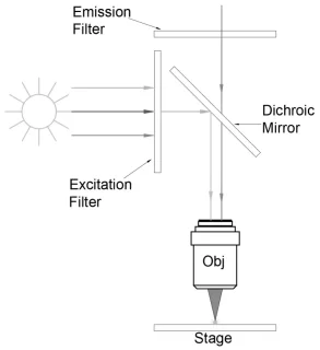
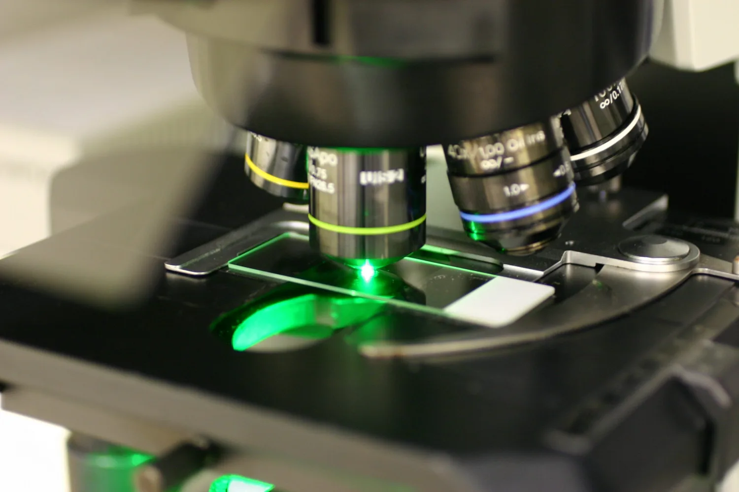
Illuminating Samples
Fluorescence microscopy, prevalent in biomedical research, exploits the phenomenon of fluorescence to visualize and study microscopic samples. Using a focused light source, such as a laser, the excitation light is directed through an excitation filter to selectively excite fluorescent molecules within the sample. A dichroic mirror separates emitted fluorescence from excitation light, ensuring only relevant signals reach the detector. The emitted fluorescence is then captured by the objective lens and directed towards a detector, allowing for the generation of detailed images.
Advantages of Fluorescence Microscopy
Selective labeling with fluorescent dyes enables the visualization of specific structures or molecules within a sample, making it invaluable for studying single-molecule interactions. Confocal fluorescence microscopy enhances depth resolution by eliminating unwanted fluorescence outside the focal plane. Automation, facilitated by computer control, enables efficient image acquisition, processing, and storage, supporting the analysis of dynamic processes such as live cell imaging.
Challenges and Future Prospects
Despite its advantages, fluorescence microscopy presents challenges, such as weak emitted fluorescence signals compared to intense excitation light. Precise optical filters are essential to eliminate unwanted background noise. Nonetheless, ongoing technological advancements and the development of new fluorescent probes continue to propel fluorescence microscopy’s pivotal role in advancing our understanding of biological systems.
Do not hesitate to contact Shanghai Optics today. We’d be more than happy to discuss your projects and how best they can become a success.