
Optics in Action: Super-Resolution Fourier Ptychographic Microscopy
Super-resolution Fourier Ptychographic Microscopy (FPM) is a modern technique that uses structured illumination, ptychography and phase retrieval to produce high resolution wide field images. With applications from metrology to drug screening, FPM provides detailed visualization with minimal aberrations.
Here we’ll look at just what the technique is about. But first, what makes it special?
Why Use Fourier Pytchographic Microscopy?
Fourier Pytchographic Microscopy (FPM) is a versatile technique that extends the capabilities of traditional microscopy in a unique way. Typically researchers would work with low magnification imaging when a wide field of view is needed, and high magnification imaging for high resolution and detailed information. But with FPM the advantages of both are combined, and a researcher can view high resolution images with fine detail over a wide field of view.
FPM also uses computational techniques to correct aberrations and improve resolution even beyond the limits of the physical microscope. This dynamic digital aberration correction and resolution enhancement is done in real time and can be adjusted as necessary.
Another benefit of FPM is the way it can extract phase information from samples under investigation. This enables even unstained transparent structures to be visible under the microscope.
What is Super-resolution Fourier Pytchographic Microscopy?
Super-resolution Fourier Pytchographic Microscopy is a computational microscopy technique that involves iteratively stitching multiple images in Fourier space to create a single image of the sample. The input images are taken with a low NA objective, and are low resolution and variably illuminated; the final image is wide field and high resolution.
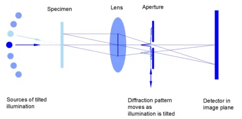
Key components of a FPM system include a low NA objective, a programmable light-emitting diode (LED) array, a digital camera or image recording device, and a series of computational reconstruction algorithms which are applied in both spatial and Fourier domains.
Applications of FPM
Super-resolution Fourier Ptyhographic Microscopy is seeing adaption in many fields, including biological research. It enables three-dimensional imaging, the reconstruction of 3D structures from 2D image data. It is also capable of label-free imaging, as it can give contrast to transparent samples even without staining fluorescent markers. A few important applications include:
Drug Screening involves determining the effect of pharmaceutical compounds on body tissue. FPM can provide the detailed images needed for a high-throughput analysis of cellular response.
Digital Pathology requires many quick, high-resolution scans of large tissue samples, and this can be easily accomplished through wide-field FPM.
But it isn’t only biological research that relies on FPM. This microscopy technique is used to provide rapid, high-precision measurements for many other scientific applications as well as for industry.
Super-resolution FPM Case Study:
The team at Shanghai Optics was asked to design a setup for super-resolution imaging of biological cells. We used a standard brightfield microscope modified with a programmed LED array, and images were reconstructed with Fourier ptychographic phase retrieval algorithms. For our initial test run, we examined Fixed HeLa cells stained with fluorescent dyes.
There were significant challenges to be overcome. The system was highly sensitive to alignment and precise calibration was needed. Preprocessing steps were required to deal with noise, and the extensive computational load meant GPU-based processing was needed.
Results, however, were impressive. We were able to achieve ~0.6 µm spatial resolution with our fast-working, affordable system. This gave us detailed visualization of sub-cellular structures, and a field of view ten times larger than that of an image acquired with confocal systems.
Although this was only a test run, our team could see immediate potential for fields like digital pathology and cancer research. The technique could be used for live imaging and might be improved by optimizing the hardware and incorporating machine learning.
Challenges to Super-Resolution FPM
Although Fourier Ptychographic Microscopy shows great promise it is not without challenges both in manufacturing and in practice. Some issues include:
- Precision: For effective FPM both the LED array and the motorized stage must have extremely accurate positioning and control, down to the nanometer level.
- Consistency: The brightness of the LED illumination must be completely uniform for all LEDs in the array.
- Minimal Aberrations: Optical aberrations must be kept to a minimum with the use of high-quality optics.
When choosing optics for a FPM setup it is important to choose components and parts from a dependable manufacturer with the capability of producing the high-precision components needed. Shanghai Optics is one example of such a manufacturer.
Advances in Super-resolution Fourier Ptychographic Microscopy
FPM research is a fast-advancing field, with strong leadership from research institutions like California Institute of Technology (Caltech), the Chinese Academy of Sciences (CAS), the University of Connecticut, Howard Hughes Medical Institute and the National Science Foundation (NSF). Recent innovations include:
- Efficient Synthetic Aperture FPM (ESA-FPM), a method that reduces the number of raw images needed to produce a super-resolution final image and, in so doing, decreases acquisition time.
- Resolution-Enhanced FPM (REFPM), a technique that incorporates advanced optical designs to produce even higher resolution images.
- Low-Rank Approximation FPM (LRA-piFP), a method that increases the robustness of the reconstruction as well as the system’s resilience to noise and environmental perturbations.
FPM at Shanghai Optics
Shanghai Optics is at the forefront of Fourier ptychographic microscopy research, and our engineers and optical designers are skilled at putting together the optimal equipment for super-resolution imaging. Contact Shanghai Optics today for more information or to place your initial order.
References:
Sun, J., Zuo, C., Zhang, L. et al. Resolution-enhanced Fourier ptychographic microscopy based on high-numerical-aperture illuminations. Sci Rep 7, 1187 https://www.nature.com/articles/s41598-017-01346-7
Zheng, G., Horstmeyer, R. & Yang, C. Wide-field, high-resolution Fourier ptychographic microscopy. Nature Photon 7, 739–745 (2013). https://www.nature.com/articles/nphoton.2013.187