All About Optical Filters for Fluorescence Microscopy
Fluorescence microscopy filters are optical filters used to selectively transmit or block specific wavelengths of light in fluorescence imaging systems. These filters improve image contrast, reduce background noise, and enable precise excitation and emission separation for fluorescence imaging applications.
Fluorescence filters are commonly used in:
- Fluorescence Microscopy
- Confocal Imaging
- Biomedical Imaging
- Raman Spectroscopy
- Life Science Research
- Analytical Instrumentation
Understanding how excitation filters, emission filters, and dichroic beamsplitters work together is essential for optimizing fluorescence imaging performance and spectral isolation.
What Is a Fluorescence Filter?
Fluorescence filters are optical filters designed to selectively transmit specific wavelengths while blocking unwanted light in fluorescence microscopy systems.
These filters help isolate fluorescence signals, improve image contrast, reduce background noise, and enhance spectral separation for high-resolution imaging applications.
A fluorescence microscope uses excitation filters, emission filters, and dichroic beamsplitters to control how light interacts with fluorophores during imaging.
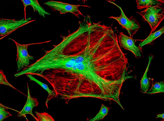
A fluorescence microscope is a type of optical microscope that uses fluorescence to examine small details or objects. Fluorescence microscopy uses optical filters to allow the transmission of certain lights through the microscope lens. This way, researchers can be able to observe specific details in objects that wouldn’t be visible to the naked eye under normal circumstances.
Different types of fluorescence filters are usually housed in a fluorescence filter cube to help enhance the visibility of small objects or details in epifluorescence microscopy, as explained below.
Types of Fluorescence Filters
Excitation Filters
Excitation filters selectively transmit wavelengths used to excite fluorophores in fluorescence microscopy systems. Modern excitation filters are typically bandpass interference filters optimized for specific fluorophore absorption spectra.
Emission Filters
Emission filters block unwanted wavelengths while transmitting fluorescence emitted by the fluorophore. These filters improve image contrast and reduce background light interference.
Dichroic Beamsplitters
Dichroic beamsplitters separate excitation and emission wavelengths by reflecting one wavelength range while transmitting another. They are typically positioned at a 45-degree angle inside fluorescence microscope systems.
Why Fluorescence Microscopy Uses Filters
Fluorescence microscopy filters help:
- Increase image contrast
- Reduce scattered light
- Block UV and IR radiation
- Improve fluorophore isolation
- Reduce background noise
- Enable multi-fluorophore imaging
- Improve spectral separation
- Enhance imaging precision
Fluorescence filter systems typically use:
- Excitation filters
- Emission filters
- Dichroic beamsplitters
- Fluorescence filter cubes
These optical components work together to isolate fluorescence signals and improve imaging quality in microscopy systems.
The first filter that light encounters in the fluorescence filter cube is the excitation filter. This filter only allows the appropriate excitation wavelength to pass through to the sample.
The transmitted light then reflects from the dichroic beamsplitter, which is typically positioned at a 45-degree angle. The dichroic filter separates excitation light from fluorescence emission light generated by the fluorophore.
Next, the emitted fluorescence passes through the emission filter, which blocks unwanted background light while transmitting the desired fluorescence wavelengths.
Finally, the fluorescence signal is directed toward the microscope eyepiece or imaging sensor, allowing researchers to capture high-resolution fluorescence images.
Fluorescence filter cubes simplify fluorescence microscopy workflows by organizing excitation filters, emission filters, and dichroic beamsplitters into a single optical assembly.
This design improves ease of use, enables rapid fluorophore switching, and allows researchers to analyze multiple fluorescence signals efficiently without repeatedly modifying the microscope setup.
Fluorescence Filter Nomenclature
Different fluorescence microscopy filters are commonly identified using abbreviated naming conventions based on their optical function and spectral characteristics.
- Excitation filters are commonly denoted as EX or X and may include ultraviolet glass (UG) or blue glass (BG).
- Emission filters are usually denoted as M or BA and may include red glass (RG) or yellow glass (GG).
- Dichroic filters are also referred to as dichroic mirrors (DM), beamsplitters (BS), chromatic beamsplitters (CBS), reflectance short pass (RKP), teiler kante (TK), or farb teiler (FB).
Conclusion
Fluorescence microscopy filters play an essential role in improving fluorescence imaging quality, spectral isolation, and signal detection in microscopy systems.
Understanding excitation filters, emission filters, and dichroic beamsplitters helps researchers optimize fluorescence imaging performance for life science research, biomedical imaging, spectroscopy, and analytical instrumentation applications.
If you’re looking for optical components, talk to us. We are manufacturers of quality and cost-effective optical components for the photonics industry. Contact us today for all your optical engineering needs.
Frequently Asked Questions
What are fluorescence microscopy filters?
Fluorescence microscopy filters selectively transmit and block specific wavelengths to improve fluorescence imaging quality.
What is the difference between excitation and emission filters?
Excitation filters illuminate the fluorophore, while emission filters isolate fluorescence emitted from the sample.
What is a dichroic beamsplitter?
A dichroic beamsplitter reflects excitation light while transmitting emitted fluorescence light.
Why are filters important in fluorescence microscopy?
Filters improve image contrast, reduce background noise, and isolate fluorescence signals.
What are fluorescence filters used for?
Fluorescence filters are used in microscopy, spectroscopy, biomedical imaging, and analytical instrumentation.