AR
CN
DEN
EN
FR
GER
ITA
POR
RUS
SPA
Optical Coherence Tomography (OCT) stands at the forefront of cutting-edge imaging technologies, revolutionizing medical diagnostics and research. Offering high-resolution cross-sectional imagery of biological tissues, OCT facilitates precise analysis and visualization of various structures, notably blood vessels.
The integration of Optical Coherence Tomography Angiography (OCTA) has further elevated the detailing and informativeness of blood vessel imaging. This article delves into the essential components and capabilities of OCT systems, their applications, and their impact on the diagnosis and management of diseases such as age-related macular degeneration (AMD) and macular edema.
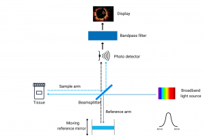
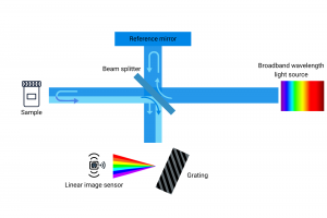
At the core of OCT technology lies low-coherence interferometry. An OCT system comprises critical elements like a light source, beamsplitter, reference arm, sample arm, and a detection system. The emitted light, often in the near-infrared spectrum, is split into optical and reference beams. The optical beam interacts with the tissue, generating interference patterns that, when processed, reconstruct cross-sectional images of the tissue.
A crucial determinant of OCT image quality is axial resolution, defining the system’s ability to distinguish structures along the depth or axial direction. Achieving resolutions in the micrometer range, OCT systems provide detailed visualization of tissue layers and microstructures.
Two primary types of OCT systems are Time-Domain OCT (TD-OCT) and Spectral-Domain OCT (SD-OCT). While TD-OCT uses a moving reference mirror for depth scanning, SD-OCT employs a spectrometer for simultaneous depth information capture, resulting in enhanced imaging speeds and signal-to-noise ratios.
OCT has evolved to include additional features like Polarization-Sensitive OCT (PS-OCT), which exploits light polarization properties to reveal information about tissue birefringence and structural characteristics. This enables visualization of collagen fibers, nerve bundles, and other polarizing tissue structures.
An extension of OCT, OCT Angiography (OCTA) facilitates non-invasive blood vessel imaging. By leveraging motion contrast of flowing blood cells, OCTA generates en-face images of the vasculature without requiring contrast agents. Analysis of changes in the OCT signal over time enables detailed mapping of the vascular network, aiding in perfusion evaluation and identification of vascular abnormalities.
OCT finds diverse applications across medical fields. In ophthalmology, it has transformed the diagnosis and management of retinal diseases, particularly AMD. High-resolution retina images allow for the identification of drusen, retinal pigment epithelial detachments, and fluid accumulation, facilitating timely diagnosis and monitoring of AMD.
The capabilities and applications of OCT continue to expand, playing a pivotal role in enhancing our understanding and management of various medical conditions. For those interested in exploring the potential of OCT in their projects, a free consultation or a quote can be requested through our contact page.
Do not hesitate to contact Shanghai Optics today. We’d be more than happy to discuss your projects and how best they can become a success.